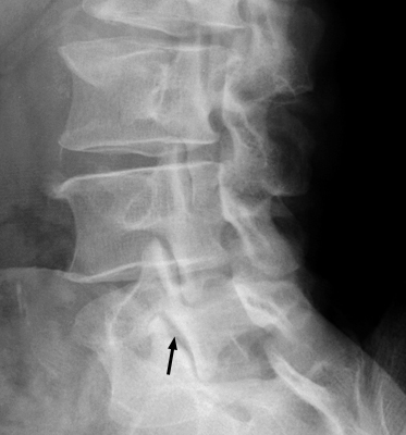
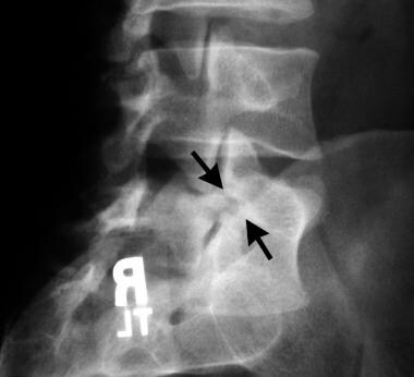
Spondylolysis (spon-dee-low-lye-sis) is defined as a defect or stress fracture in the pars interarticularis of the vertebral arch. the vast majority of cases occur in the lower lumbar vertebrae (l5), but spondylolysis may also occur in the cervical vertebrae.. Fig. 1: oblique x-ray view of the lumbar spine ("scotty dog" view). in the lower segment, a fracture (or spondylolysis) through the pars shows up as a black collar on the dog's neck. interestingly though, with radiological investigation, occasionally no fracture is seen, but presents as an elongated pars interarticularis.. When viewing the lumbar spine in a anterior oblique view the result is an image which resembles a "scotty dog". a fracture of the par interarticularis (isthmus) results on the x-ray as a dark "collar" on the neck of the dog (#3 on the graphic)..
Modality: x-ray used in the following article: scottie dog sign (spine) - “ the scottie dog sign refers to the normal appearance of the lumbar spine when seen on oblique radiographic projection.. The scotty dog sign refers to the normal appearance of the lumbar spine when seen on oblique radiographic projection. on oblique views, the posterior elements of vertebra form the figure of a. Oblique projection radiograph shows the presence of bilateral pars defects (arrows), with an appearance resembling a scottie dog with a collar. (the collar is the pars defect.) spondylolisthesis..


0 komentar:
Posting Komentar